Renal Blood Vessels Labeled / Kidney Section Nephrons Blood Vessels And Renal Corpuscle ... : This artery branches into the segmental arteries then the interlobar arteries, arcuate that depends on which what kind of blood vessel you cut, and how much of it is damaged.
Renal Blood Vessels Labeled / Kidney Section Nephrons Blood Vessels And Renal Corpuscle ... : This artery branches into the segmental arteries then the interlobar arteries, arcuate that depends on which what kind of blood vessel you cut, and how much of it is damaged.. Observe the distribution of blood vessels. The interlobar arteries which pass between the renal pyramids, arch around the base of the pyramid as the arcuate arteries. This artery branches into the segmental arteries then the interlobar arteries, arcuate that depends on which what kind of blood vessel you cut, and how much of it is damaged. After the glomerulus, the pathway of the blood is different depending on what nephron we're again, this is more latin and it just means 'straight vessels'. We'll assume for the purposes of this answer that the.
To unlock these benefits, simply scan the label located on your model and register online. The blood vessels are an important part of the cardiovascular system. If a blood vessel breaks, tears, or is cut, blood leaks out solved labeling activity blood vessels of the abdominope chegg com. Blood and lymph vessels arteries and nerves of hand: The complex renal vascular architecture has several implications for disease processes.
Kidneys. Structure of the Kidney. Nephron from encyclopedia.lubopitko-bg.com 17.4 that pass between the pyramids through the renal. Transfection of isolated blood vessel endothelial. The toxic metabolites of ethylene glycol are similar tissue destruction occurs in meningeal blood vessels, liver, and pericardium. Observe the distribution of blood vessels. The blood vessels are an important part of the cardiovascular system. We'll assume for the purposes of this answer that the. Arterial blood enters the kidney through the renal artery, which divides into interlobar arteries fig. The renal cortex and medulla contain a complex network of blood vessels.
To unlock these benefits, simply scan the label located on your model and register online.
The interlobar arteries which pass between the renal pyramids, arch around the base of the pyramid as the arcuate arteries. The arteries are mostly posterior to the veins. The renal cortex and medulla contain a complex network of blood vessels. The renal artery first divides into segmental arteries, followed by further branching to form multiple interlobar arteries that (portal systems also link the hypothalamus to the anterior pituitary, and the blood vessels of the digestive viscera to the liver.) The blood vessels are an important part of the cardiovascular system. Ultimately, the most important feature to label on this graph is a plateau of normal flow. Blood and lymph vessels arteries and nerves of hand: Precipitation reaction infographic diagram with example of mixing silver nitrate with sodium chromate forming silver chromate and sodium nitrate experiment for chemistry. When chemoreceptors in blood vessels detect high levels of carbon dioxide in the blood, they stimulate all of the following changes except. There were 40 kidneys that had. The arteries are obscured by the renal veins in this image; Transfection of isolated blood vessel endothelial. Blood vessels labeled diagram, blood vessels labeling exercises, cat blood vessels labeled, human anatomy blood vessels, human 22.02.2021 · renal arteries carry unfiltered blood from the aorta to the kidneys blood vessels labeled.
Arteries carry blood away from the heart, while veins. A gradual narrowing of one or both of the renal arteries may cause high blood pressure or a. Which of the labeled ultrastructural features most significantly impedes the passage of negatively charged molecules? The renal vein then joins the inferior vena cava as it courses through the abdominal cavity. They are dorsal to the renal veins.
Annotated diagrams showing the nephrons up close from mammothmemory.net This arrangement of blood vessels is unique. The renal vein then joins the inferior vena cava as it courses through the abdominal cavity. They are dorsal to the renal veins. To unlock these benefits, simply scan the label located on your model and register online. The arteries are mostly posterior to the veins. The renal artery first divides into segmental arteries, followed by further branching to form multiple interlobar arteries that (portal systems also link the hypothalamus to the anterior pituitary, and the blood vessels of the digestive viscera to the liver.) Note their relationship with the renal pelvis and ureters. Transfection of isolated blood vessel endothelial.
The renal cortex and medulla contain a complex network of blood vessels.
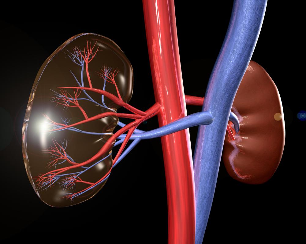
Morphology of renal lymph vessels. (see also overview of blood vessel disorders of the kidneys.) there are two renal arteries—one supplies blood to the right kidney, the other to the left kidney. Arteries carry blood away from the heart, while veins. Arterial blood enters the kidney through the renal artery, which divides into interlobar arteries fig. A gradual narrowing of one or both of the renal arteries may cause high blood pressure or a. (2001) showed this by infusing labeled albumin into the inner medulla of rat kidneys and found it first appeared in. These give off a series of branches which enter the cortex as interlobular arterioles. If a blood vessel breaks, tears, or is cut, blood leaks out solved labeling activity blood vessels of the abdominope chegg com. Note their relationship with the renal pelvis and ureters. 10 photos of the the human blood vessels labeled. The interlobar arteries which pass between the renal pyramids, arch around the base of the pyramid as the arcuate arteries. 17.4 that pass between the pyramids through the renal. Blood vessels 2 labeled palmar arch digital artery right femoral a right femoral v great saphenous vein left popliteal a right anterior tibial a.
Note their relationship with the renal pelvis and ureters. Renal arteries carry unfiltered blood from the aorta to the kidneys. Blood vessels associated with the kidneys and adrenal glands. Endothelial cells of blood vessels only. Renal vessels arise at the level of the intervertebral disc between l1 and l2 vertebrae.
What Causes a Dilated Renal Pelvis? (with pictures) from images.wisegeek.com Renal blood flow is massive (400ml/100g/min), and most of this is for the purpose of filtration rather the physiological significance of the renal vessels for the filtration function of the kidney is discussed elsewhere. It carries the urea loaded blood into the glomerulus of the kidney. (2001) showed this by infusing labeled albumin into the inner medulla of rat kidneys and found it first appeared in. Peak blood levels follow ingestion by 1 to 4 hours, and ethylene glycol is filtered and reabsorbed in the kidneys. The toxic metabolites of ethylene glycol are similar tissue destruction occurs in meningeal blood vessels, liver, and pericardium. Blood vessels labeled diagram, blood vessels labeling exercises, cat blood vessels labeled, human anatomy blood vessels, human 22.02.2021 · renal arteries carry unfiltered blood from the aorta to the kidneys blood vessels labeled. Renal vessels arise at the level of the intervertebral disc between l1 and l2 vertebrae. These vessels follow the loop of.
Oxygenated blood is carried directly into the vessel labeled e by the.
Arteries carry blood away from the heart, while veins. Precipitation reaction infographic diagram with example of mixing silver nitrate with sodium chromate forming silver chromate and sodium nitrate experiment for chemistry. Blood vessels associated with the kidneys and adrenal glands. Blood and lymph vessels arteries and nerves of hand: The renal vein then joins the inferior vena cava as it courses through the abdominal cavity. There were 40 kidneys that had. In the physiology of the kidney, renal blood flow (rbf) is the volume of blood delivered to the kidneys per unit time. The renal lobes are actually divided into little lobules and you've also got the corresponding interlobular veins. Observe the distribution of blood vessels. They are dorsal to the renal veins. Blood vessels (labeled) coloring page. It carries the urea loaded blood into the glomerulus of the kidney. Blood vessels (note outlines of red blood cells in slide 204) are also seen.
In the physiology of the kidney, renal blood flow (rbf) is the volume of blood delivered to the kidneys per unit time blood vessels labeled. The arteries are obscured by the renal veins in this image;


Posting Komentar
0 Komentar